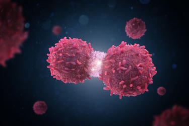
3D Cell Culture Applications And Bioprinting Will Change How We Research Cancers

2D cell culture and animal models have led to major discoveries and improved understanding of many of these diseases, but they have their limitations. With the advent of 3D cell culture applications and 3D bioprinting, cancer research is changing. Now, complex aspects of cancer like metastasis can be modeled and studied, and the efficacy and toxicity of drugs can be tested more realistically and rapidly. Cells grown in 3D conditions more accurately mimic in vivo cellular responses, according to a recent study in Annals of Biomedical Engineering.
Different 3D bioprinting approaches have investigated specific aspects of tumor biology and drug testing. 3D printed matrices are "ideal platforms for the creation of 3D tumor spheroids possessing hypoxic cores," according to a study in Current Opinion in Biomedical Engineering.
In recent years, 3D cell culture applications have developed rapidly, allowing for the creation of more realistic biochemical microenvironments in which to study cancer and its response to therapy. As techniques and systems continue to evolve and develop, so too will our understanding of cancer and the selection of new therapeutic targets for chemotherapies.
In this blog we explore how 3D bioprinting works and how it is advancing cancer research.
Get unlimited access to:
Enter your credentials below to log in. Not yet a member of Cell & Gene? Subscribe today.
